A patient comes in having been told at another clinic that they need an implant, sometimes two or three, and that the procedure is straightforward. No CBCT was taken. The treatment plan was built from a flat panoramic X-ray and a visual examination.
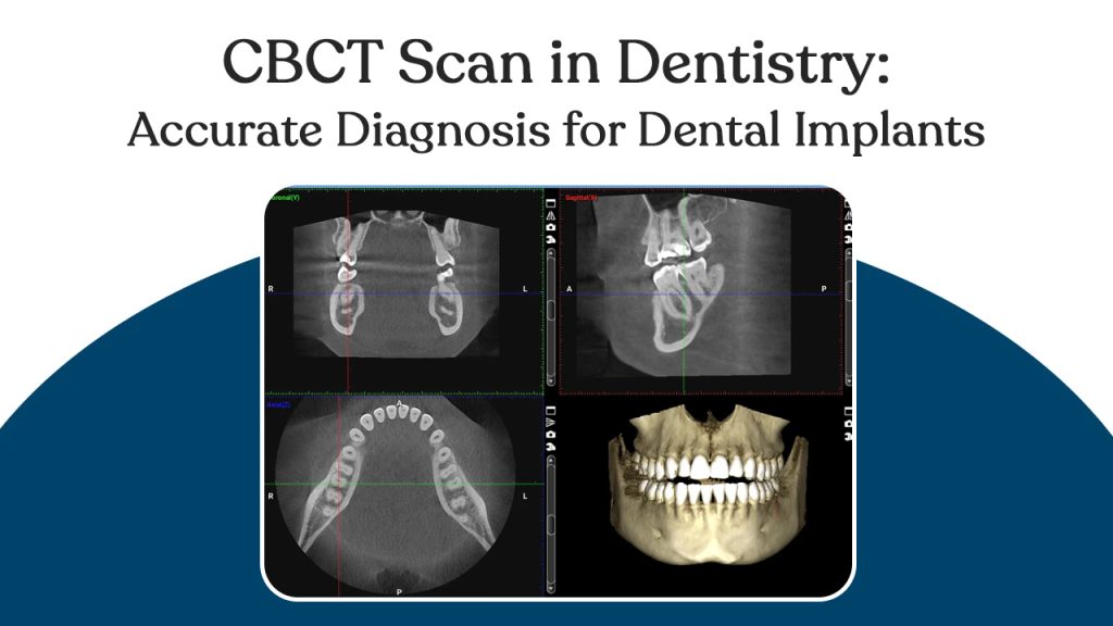
A CBCT scan in dentistry creates a three-dimensional map of the entire surgical site and any hidden pathology like cysts or retained root fragments. That map is what accurate implant diagnosis is built from. Without it, the surgical plan is built on partial information.
At Asian Dental, the super-speciality implantology team performs CBCT imaging as a standard for every implant case. Over 1,000 implants were placed using gold-standard instruments, laser technology, 4-step WHO sterilisation protocol. Thus the CBCT scan for dental implants is not an optional add-on at Asian Dental. It is where the implant plan begins.
What Is a CBCT Scan and Why Is It Not the Same as a Regular Dental X-Ray?
Patients often ask why a standard X-ray is not sufficient and CBCT is. The answer is in the dimension of information each one produces and what that dimension difference means for surgical planning.
A conventional dental X-ray produces a two-dimensional flat image of a three-dimensional structure. It shows tooth position, root length, and a general bone outline.
What it cannot show: bone width front to back, precise density at any point, the exact three-dimensional nerve path through the lower jaw, or the true sinus floor height above upper back teeth.
A CBCT rotates around the patient’s head in a single pass, capturing hundreds of images from different angles. Software assembles these into a full three-dimensional model of the jaw. Every measurement that was an estimate becomes a precise number.
| Feature | Conventional X-Ray | CBCT Scan |
| Image dimension | 2D flat image | Full 3D volumetric model |
| Bone width measurement | Cannot measure | Precise to the millimetre |
| Bone density assessment | Approximate | Classified by density grade |
| Nerve location | Approximate 2D position | Exact 3D pathway mapped |
| Sinus floor height | Estimated | Measured precisely |
| Hidden pathology detection | Limited | Cysts, infections, root fragments visible |
| Radiation dose | Low | Very low — 10x less than medical CT |
| Scan duration | Seconds | Under 60 seconds |
How CBCT Makes Dental Implant Diagnosis Accurate?
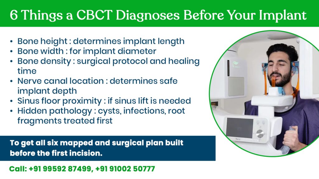
At a dental hospital in Hyderabad, the CBCT report is what every implant consultation is built around. Here are the six clinical data points the scan provides and what each one changes about the diagnosis and surgical plan.
- Bone height: The vertical distance from the ridge crest to the nerve canal below (lower jaw) or sinus floor above (upper back teeth). Standard implants require a minimum of 10mm. Below that, the plan changes shorter implants, sinus lift, or nerve repositioning. The panoramic X-ray gives an approximation. The CBCT gives a number.
- Bone width: The horizontal thickness of bone at the planned implant depth. An implant needs a minimum of 1mm of bone on each side for stable integration. Bone that looks adequate in height on a 2D X-ray is sometimes too narrow in width. The CBCT reveals this before surgery.
- Bone density: Classified from Type I (dense cortical) to Type IV (soft, largely cancellous). Type IV bone requires a different surgical protocol, different implant design, and longer healing before loading. Without density data, the surgeon discovers this at the moment of placement.
- Nerve location: The inferior alveolar nerve runs through the lower jaw in a canal. Contact during placement causes numbness of the lower lip and chin. The CBCT by the best dentist in Kondapur maps the nerve’s three-dimensional path, allowing the surgeon to plan depth with a documented safety margin.
- Sinus floor proximity: The maxillary sinuses sit above the upper back teeth. Insufficient height means an implant can perforate the sinus, a serious complication. The CBCT measures the exact distance.
- Hidden pathology: Retained root fragments, periapical cysts, early-stage infections, and areas of bone loss invisible on a panoramic X-ray appear clearly on CBCT.
What Happens When Implants Are Placed Without an Accurate CBCT Diagnosis?
Patients comparing implant providers sometimes find clinics that offer treatment without a CBCT, substituting a panoramic X-ray or proceeding from clinical examination alone. Here is what that means in practice.
- Nerve damage is the most serious risk. The inferior alveolar nerve cannot be precisely located on a 2D image.
- Sinus perforation follows the same logic in upper jaw cases. Without measuring the exact distance to the sinus floor, the surgeon is estimating clearance.
- Wrong implant dimensions are selected when bone width is unknown. An implant placed into bone too narrow for it fails under load.
- Bone grafting discovered mid-surgery is an informed consent failure. A patient who consented to a single-stage procedure and is told mid-appointment that grafting is required did not have accurate diagnostic information going in.
From Accurate Diagnosis to Placed Implant: How CBCT Data Builds the Surgical Plan at Asian Dental?
The CBCT scan at Asian Dental, a dental clinic in Kondapur feeds directly into the surgical planning workflow and for eligible cases, eliminates the need for a conventional incision entirely.
After the CBCT scan in dentistry, the implantologist plans implant position, angle, and depth digitally on the three-dimensional model. Every measurement is confirmed against the anatomical landmarks the scan has mapped. Nerve safety margins documented. Sinus clearance confirmed. Bone density at planned depth classified.
For cases where anatomy supports it, a surgical guide precision-milled from the CBCT data is produced before the surgical appointment. The guide fits over the patient’s teeth and controls drill direction and depth during placement.
Accurate diagnosis for dental implants hyderabad is not just better imaging. It is a surgical workflow that is planned, verified, and executable precisely because nothing was estimated.
Call to book an appointment:
Kondapur No. +91- 99592 87499
Kukatpally No. +91- 91002 50777
Frequently Asked Questions
Is a CBCT scan painful or safe for patients?
A CBCT scan is completely painless, you sit or stand while the machine rotates around your head for under sixty seconds. No injection, no contact with the mouth, no preparation needed..
Is a CBCT scan necessary for every dental implant?
For single implants in patients with known adequate bone and no complicating anatomy, some surgeons use panoramic X-rays. At Asian Dental, CBCT is standard for every implant case because the nerve location, bone density, and sinus proximity data it provides is what the surgical plan is built from.
How long does a CBCT scan take at the dentist?
The scan itself takes under sixty seconds. The full appointment including positioning, scan, and initial image review runs fifteen to twenty minutes.
Can a regular X-ray replace a CBCT scan for implant planning?
No. A panoramic or periapical X-ray provides a two-dimensional image that cannot measure bone width, classify bone density, or map the precise three-dimensional path of the inferior alveolar nerve.


